Plasma needle investigations:
We built a low-power millimeter-size atmospheric plasma jet and tested it for dental applications.
The apparatus is modeled after the "plasma needle" introduced by Eva Stoffels and her group in Holland.
Teaming with Professor David Drake of the The University of Iowa College of Dentistry, we performed tests to demonstrate that the plasma needle kills Streptococcus mutans (S. mutans) bacteria, which is the most important microorganism for causing caries (cavities).
Papers we've published can be found here
Images from July 2005 tests:

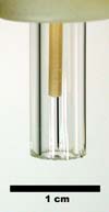
Nozzle of the plasma needle handset. Helium gas flows from this nozzle,
and radio-frequency high voltage is applied to the tungsten needle to
ionize the gas. A plasma jet flows out of the nozzle and mixes with air.
Radicals O and OH are formed when air molecules are dissociated. The plasma
jet impinges on a surface.

We imaged the glow from the side and then used Abel inversion to reveal
its internal structure. The needle is just above the center of the image,
while the treated surface is horizontal, at the bottom.

Plasma treatment was performed in 501 Van Allen Hall by John Goree (left)
and Bin Liu. The samples were prepared, incubated, and imaged the Dental
Science Building by the group of Dr. David Drake.

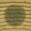
Image of a 5-mm diameter plasma-treated spot on the surface of an agar
plate. A light-brown color indicates living bacterial colonies, while
a dark-brown color indicates dead bacteria. This serves not only as an
indication of a desired biomedical effect, but also as a sensitive biological
diagnostic of plasmas.
